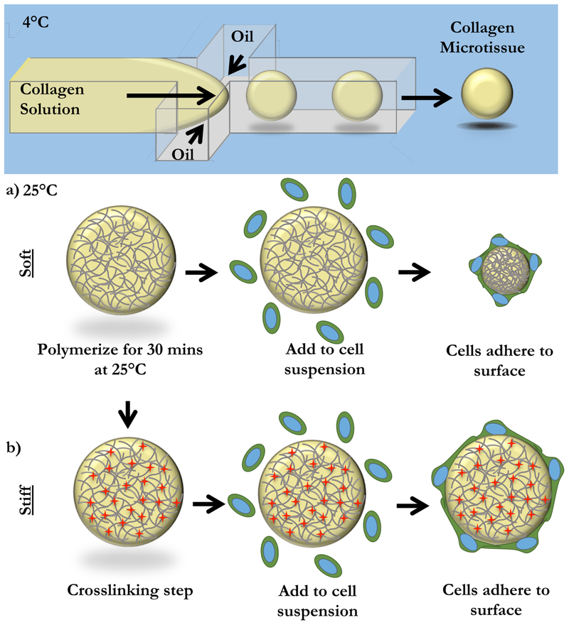
Figure 1: Fabrication of Endothelial-coated Collagen Microtissues.
Liquid 6 mg/mL collagen I microtissues were generated using a chilled flow-focusing microfluidic device. Microtissues were collected and polymerized off-chip at 25°C for 30 minutes. (a) In the case of “soft” microtissues, we did not need to further manipulate the collagen microtissues. To coat with cells, microtissues were mixed with a single-cell suspension of endothelial cells. In this “soft” mode, cells compacted the collagen microtissues. (b) To prevent compaction, we created “stiff” microtissues. After polymerization, microtissues were crosslinked with formalin. “Stiff” microtissues were then washed thoroughly and coated by incubating with a single cell suspension of endothelial cells. Cells created a monolayer on the surface of the microtissues, but did not change the overall size of these “stiff” microtissues.

