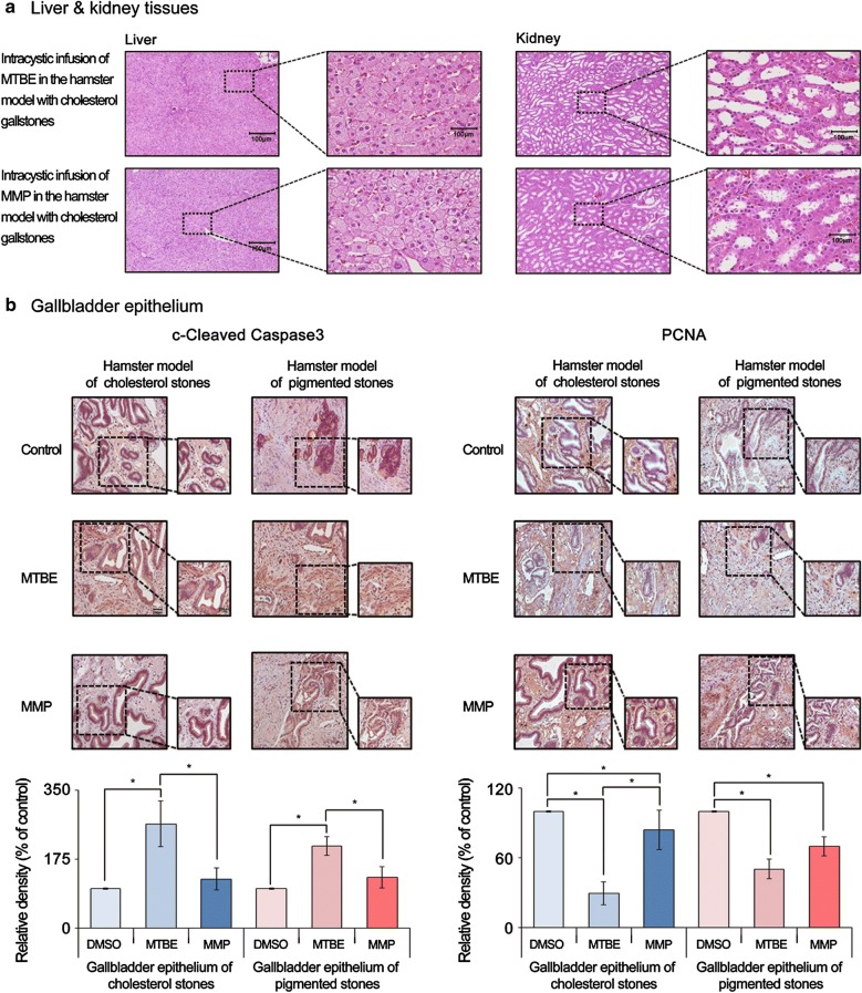
Fig. 5.
Validation of tissue toxicities of each gallstone-dissolving compound in hamster models of gallstones. a Hematoxylin–Eosin (H&E) stains of the liver and kidney tissues obtained from the gallstone-containing hamsters at 24 h after intracystic injection of each solvent. In HE stains, the liver and kidney tissues of each group appeared to be well-preserved and did not demonstrate any signs of injury. b [Left] Cleaved caspase-3 immunohistochemical stains of the gallbladder tissues obtained from the hamsters with gallstones at 24 h after intracystic injection of each solvent. Whereas MTBE significantly increased the expression of cleaved caspase-9 (P < 0.05), MMP did not in both hamster models of cholesterol and pigmented gallstones. [Right] PCNA immunohistochemical stains of the gallbladder tissues obtained from the hamsters with gallstones at 24 h after intracystic injection of each solvent. Although both solvents significantly decreased the expression of PCNA, the reduction in PCNA expression was far significant after MTBE than it was after MMP in both gallstone models (P < 0.05). *P < 0.05. DMSO, dimethyl sulfoxide; MMP, 2-methoxy-6-methylpyridine; MTBE, methyl tert-butyl ether

