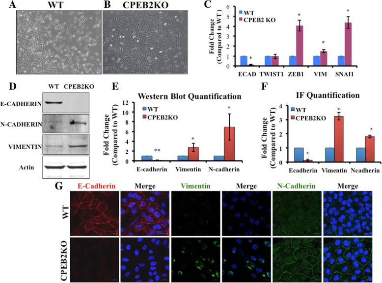
Fig. 2.
Epithelial to mesenchymal transition in CPEB2KO cells. a Wildtype (WT) MCF10A cells exhibiting epithelial cell morphology. b CPEB2KO cells exhibiting elongated, mesenchymal (Fibroblast-like) morphology (Magnification viewed at10x objective). c Quantitative RT-PCR for EMT marker mRNAs in WT and CPEB2KO MCF10A cells. Epithelial marker E-Cadherin was significantly decreased (to 0.116 fold), with significant increases in mesenchymal markers ZEB1 (4.06 fold), Vimentin (1.49 fold), and SNAI1 (4.38 fold) in CPEB2KO cells. d, e Induction of EMT in CPEB2KO cells shown at the protein level. Representative Western blots (d) and quantification of Western Blots (e) for E-Cadherin, Vimentin and N-Cadherin in wildtype and CPEB2KO cell lines showing significantly decreased E-Cadherin protein (to 0.078 fold), significantly increased Vimentin (to 2.75 fold) and N-Cadherin (to 6.93 fold) in CPEB2KO cells. f, g EMT visualized through immunofluorescence of markers. g. E-Cadherin (red), Vimentin (green), and N-Cadherin (green); Nuclei (blue). Magnification Scale = 20 μm. f. Integrated fluorescence density quantified with ImageJ Software and normalized to cell number, showing that E-Cadherin protein expression was significantly decreased (to 0.20 fold), with a significant increase in Vimentin (to 3.50 fold) and N-Cadherin (to 1.80 fold) in CPEB2KO cells compared to WT cells. Data presented as mean of 3 replicates ± SEM. (*) indicates p < 0.05, (**) p < 0.001

