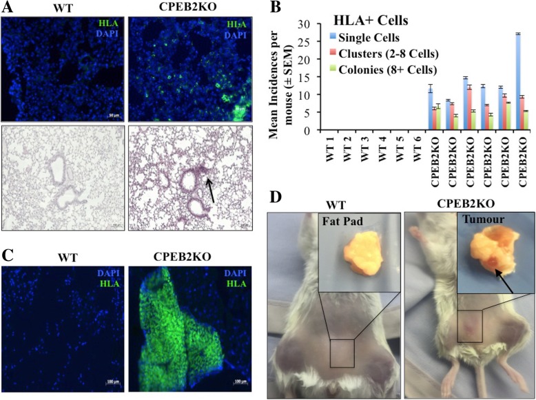
Fig. 7.
Tumorigenicity of WT and CBEB2KO MCF10A cells in NOD/SCID/IL2Rγ null mice. a, b Intravenous injection of cells (5 × 10⁵ cells per mouse, n = 6 mice per cell line) resulted in lung metastasis of CPEB2KO but not WT cells. a Top panel: HLA stained (green) tumor cells (single cells, clusters and colonies) noted in the lungs with CPEB2KO inocula. Nuclei stained blue with DAPI. Bottom panel: H&E stained lung showing a small tumor-like lesion (pointed with arrow) with CPEB2KO inoculam. Scale = 50 μm (IF images) and 100 μm (H&E images). b Incidence of single tumor cells, clusters (2–8 cells) and colonies (more than 8 cells) within lung sections immunostained with HLA antibody. Tumor cells were identified in the lungs of all mice inoculated with CPEB2KO cells but none of the lungs in mice inoculated with WT cells. Data presented as mean of 5 images per section, 3 non-serial sections per mouse ± SEM. c, d Subcutaneous inocula (5 × 10⁵ cells per site, 2 inguinal mammary sites per mouse, mixed with Matrigel, n = 5 mice in each group) of WT and CPEB2KO cells at the mammary sites of NOD/SCID/IL2Rγ-null mice. CPEB2KO cells formed local tumours in all mice, some of which (2 out 5, 40%) spontaneously metastasized to the lung. No tumor resulted in any mouse from WT cells. Tumour-forming efficiency was 90%, as calculated by number of sites showing local tumours (nine) divided by total number of injection sites (ten). Each mouse showed one or two tumors: 4 with double and 1 with single tumor. d Representative images of mice and mammary fat pads at 12 weeks. Arrow pointing to a tumor. c Representative images of tissues from sites inoculated with WT or CPEB2KO MCF10A cells, immunostained with HLA antibody. Tumor cells (identified only with CPEB2KO cells) stained green, and nuclei stained blue with DAPI

