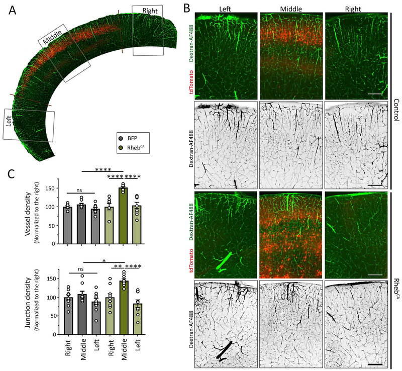
Figure 3: Hypervascularization is restricted to the MCD.
(A) Low magnification image of the cortex containing the tdTomato-fluorescent electroporated region and dextran-AF488 fluorescence vessels. (B) Images of tdTomato-positive cells containing a control plasmid (noted control) or RhebCA (noted in the right) and dextran-AF488-filled blood vessels in green or in black (shown alone) in coronal sections from the somatosensory cortex. Scale bars: 200 μm. (C) Bar graphs of vessel density and normalized junction density in control (grey) and RhebCA (green) conditions. N≥2 sections per mouse from 4-5 mice (datapoints on graph= value per section). Two-way ANOVA followed by Tukey test, ****: P<0.0001, **:P<0.01, *:P<0.05, and ns: not significant.

