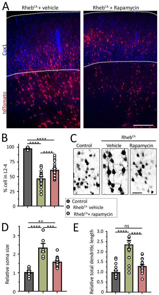
Figure 4: Rapamycin partially rescues neuronal defects in focal MCDs. (A Figure 4: Rapamycin partially rescues neuronal defects in focal MCDs.
(A) Images of tdTomato-positive RhebCA-expressing neurons and Cux1 immunostaining in coronal sections from mice electroporated with RhebCA at E15 and repeated with either vehicle or 0.5 mg/kg rapamycin from P1 to P14 every other day. A section containing control BFP-electroporated neurons is shown in Figure 2. Scale bars: 300 μm. (B) Bar graphs of the percentage (%) of tdTomato+ control neurons (grey) and RhebCA-expressing neurons in L2-4 (based on Cux1 staining) in mice treated with vehicle (green) or rapamycin (red). (C) Images of control neurons in mice electroporated with BFP + tdTomato and RhebCA-expressing neurons in mice electroporated with RhebCA + tdTomato and treated with vehicle or rapamycin. (D and E) Bar graphs of the soma size and total dendritic length of control neurons (grey) and RhebCA neurons in mice treated with vehicle (green) or rapamycin (red). N>3 mice, 2 sections per mouse (each datapoint is the mean from >8 neurons per section for dendrite analysis and >12 neurons for cell size per section). One-way ANOVA with Tukey post-test. ****: P<0.0001, **:P<0.01, *:<P0.05, ns: not significant.

