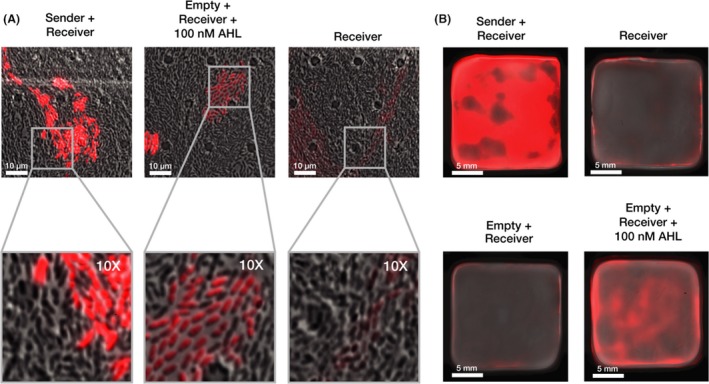
Figure 3.

Cell‐to‐cell signalling functions within co‐cultured pellicles.A. Fluorescence microscopy of co‐cultures. Images detail K. rhaeticus growth after 48 h within three separate chambers on the same microfluidics plate. K. rhaeticus co‐cultures were produced from a 1:1 inoculation of each strain. Images were taken with a 60× oil emulsion lens. The bottom column displays a digitally zoomed in region of the images in the top. All microscopy images were taken with the same settings for brightfield and mRFP fluorescence channels. Brightness and contrast for the brightfield channel were adjusted to improve clarity, while the red fluorescence channel was left unadjusted (B) Pellicle co‐cultures. The top left pellicle is a 1:1 mix of Sender and Receiver culture and the top right a pure Receiver pellicle. Both the bottom pellicles are a 1:1 mix of Empty and Receiver, where the bottom left was left to grow without AHL while the bottom right had 100 nM AHL added to it on second day of growth.
