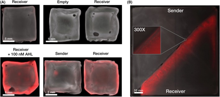
Figure 4.

Cell‐to‐cell signalling can function between separate Sender and Receiver pellicles. All pellicle images have two colour channels, a pseudo‐brightfield and red fluorescence channel.
A. Receiver pellicle induction by Sender pellicle. Left images show two Receiver pellicles after 24 h incubation in 5 ml HS media, the top pellicle without any added AHL and the bottom pellicle with 100 nM AHL. The right images show Receiver‐Sender and Empty‐Sender pellicle pairs after 24 h co‐incubation in 5 ml HS media.
B. Boundary detection. Spliced Sender‐Receiver pellicle was grown for 24 h and then removed from soft agar surface and imaged. A digitally zoomed in region of the boundary between the two pellicles is featured.
