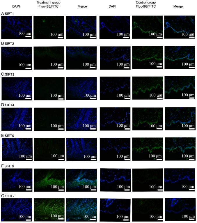
Figure 6.
IF in mouse skin lesions. IF in mouse skin lesions were also negative for (A) SIRT1, (B) SIRT2, (C) SIRT3, (D) SIRT4 and (E) SIRT5 antibodies. Lesions were positive for (F) SIRT6 and (G) SIRT7 antibodies. SIRTs were mainly localized in the epithelial layer. IF, immunofluorescence; SIRT, Sirtuin; FITC, fluorescein isothiocyanate.

