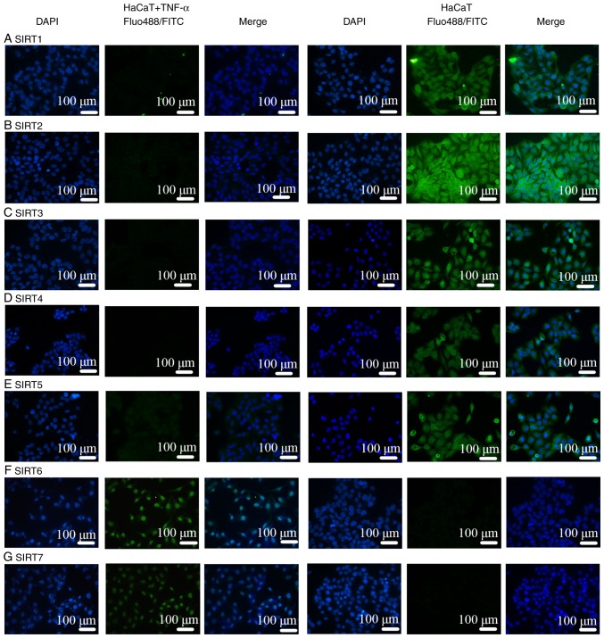
Figure 7.
IF in TNF-α-stimulated HaCaT cells was negative except for SIRT6 and 7. (A) SIRT1 and (B) SIRT2 were located predominantly in the nucleus and cytoplasm. (C) SIRT3, (D) SIRT4 and (E) SIRT5 were primarily mitochondrial proteins. (F) SIRT6 and (G) SIRT7 were mainly nuclear Sirtuins. IF, immunofluorescence; SIRT, Sirtuin; FITC, fluorescein isothiocyanate.

