Figure 1.
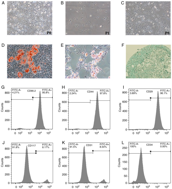
Identification of primary BM-MSCs. Primary BM-MSCs were isolated from the femur and tibia of 4-week-old C57BL/6 mice. (A-C) Morphology of BM-MSCs was observed under light microscopy at P0, P1 and P5 (magnification, ×100). (D-F) Differentiation potentials of BM-MSCs into adipocytes, osteoblasts and chondrocytes were confirmed with Oil Red O staining (magnification, ×200), Alizarin Red S staining (magnification, ×100) and Alcian blue staining (magnification, ×400), respectively. (G-L) Flow cytometric detection of surface markers on BM-MSCs. Cells were positive for CD90.2, CD44 and CD29, and negative for CD117, CD31, and CD34. BM-MSCs, bone marrow-derived mesenchymal stromal cells; CD, cluster of differentiation; FITC, fluorescein isothiocyanate; P, passage.

