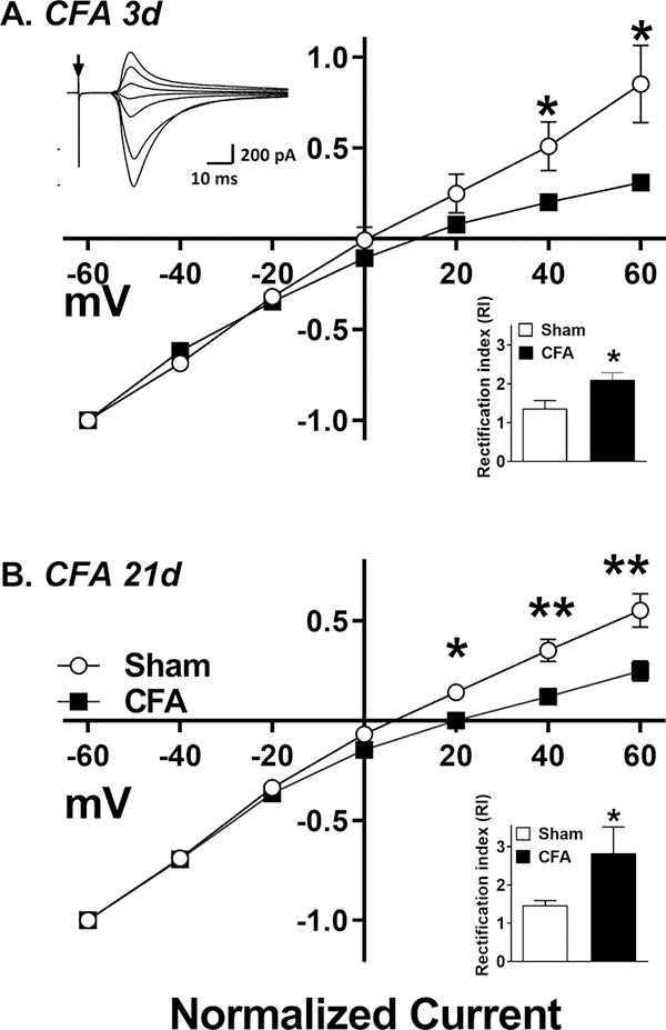
Figure 1. AMPA receptors retain Ca2+ permeability in CFA 21d mice.
EPSCs were recorded at different membrane potentials from −60 mV to +60 mV in 20 mV steps. The I-V curves represent the C-fiber mediated, evoked peak EPSCs measured at (A) 3 d and (B) 21 d after CFA injury. Inset, top-left; example plot of AMPAR mediated EPSCs at various membrane potentials. Arrow marks time of DRS. Inset, bottom-right; RI for saline vs. CFA treated animals calculated as described in methods. Data represent mean ± SEM. N = 5–6 mice/group. *p<0.05, **p<0.01 vs sham group.

