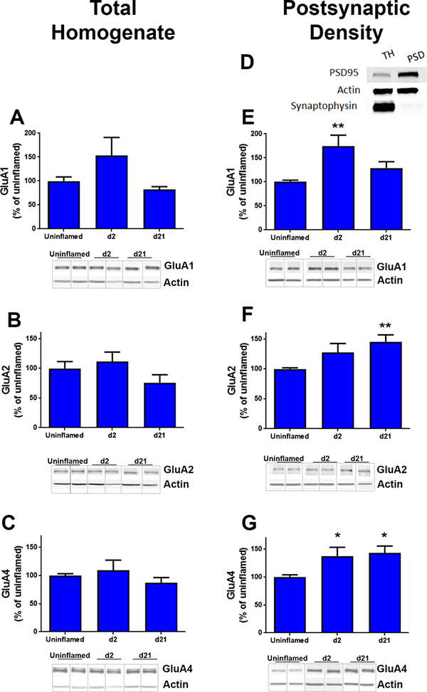
Figure 2. Inflammation increases the expression of GluA1, GluA2 and GluA4 subunits in PSD.
Western blots were performed using the following primary antibodies; (A, E) anti-GluA1, (B, F) anti-GluA2, and (C, G) anti-GluA4. Representative blots are shown below each graph. D. Subcellular fractionation: A representative western blot shows enrichment of PSD-95 (postsynaptic marker) and the absence of synaptophysin-I (presynaptic marker) in postsynaptic density fractions from dorsal horns. Densitometry was performed to quantify pixel density in each western as a measure of subunit expression at d0, d2 and d21 post-CFA injection. Quantification was performed relative to β-actin levels. Ipsilateral dorsal horns from lumbar L3/L4 spinal cords of four mice were pooled to obtain each individual sample. * p<0.05 n= 4–7 samples per time point. All blots and data analyses are available in Supplemental Figure 1.

