
Figure 2. Massively increased Hsp90α in wound bed following skin injury in mice.
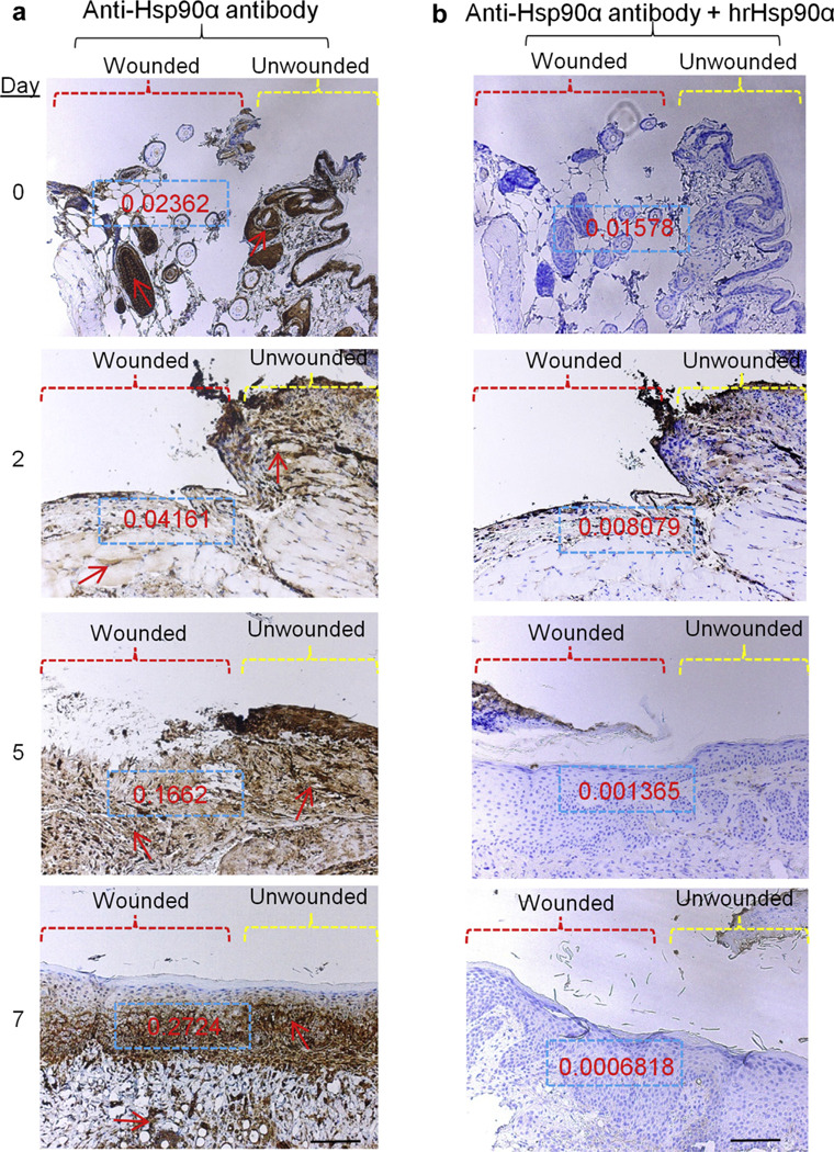
Nine 0.8 cm × 0.8 cm full-thickness excision wounds were created on the back of the mice.The wounds were monitored every 24 hours and wedge biopsies of partial (day 0 in particular) or entire wounds were taken on indicated days. Sections of the wounds were subjected to immunohistochemical analyses, exactly as described in Figure 1. These data represent a consensus from multiple and noncontinuous sections of skin specimens. Scale bar = 1.5 mm.Hsp, heat shock protein.