
Figure 5. Topically applied recombinant Hsp90α-Δ protein promotes wound closure as effectively as Hsp90α-wt protein.
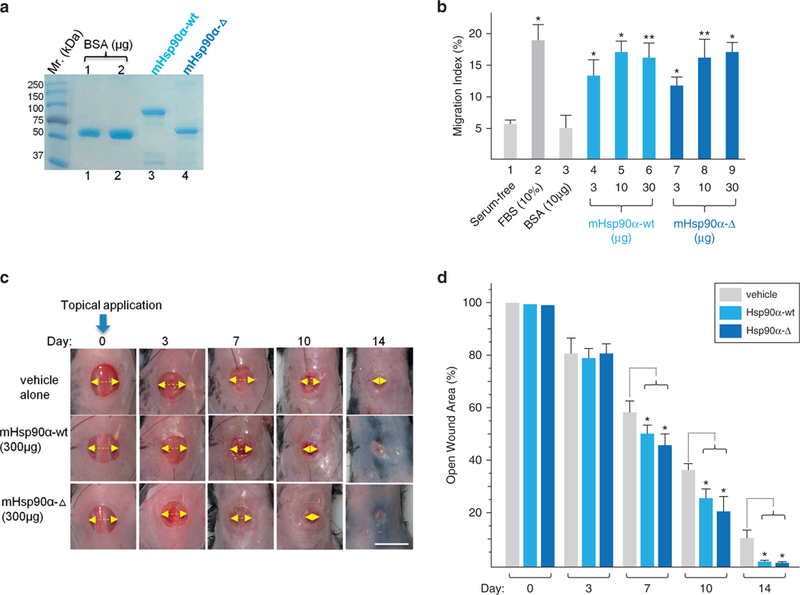
(a) Recombinant mouse Hsp90α-Δ and mouse Hsp90α-wt proteins were produced from cDNAs isolated from the dermal fibroblasts of the mice by pET bacterium inducible expression system, purified by fast protein liquid chromatography and visualized on a Coomassie blue-stained SDS gel. (b) Colloidal gold migration assay of human keratinocytes under the indicated stimulations. (c) Images of wound closure in Hsp90α-wt mice under carboxymethyl cellulose, Hsp90α-Δ, or Hsp90α-wt protein treatment. The yellow arrow indicates the relative distance of the yet unhealed portion of a wound. (d) Quantitation of the triplicate wounds from the experiment shown in (c), n = 3. *P ≤ 0.05. Scale bar = 1.0 cm. FBS, fetal bovine serum; Hsp, heat shock protein; wt, wild type.