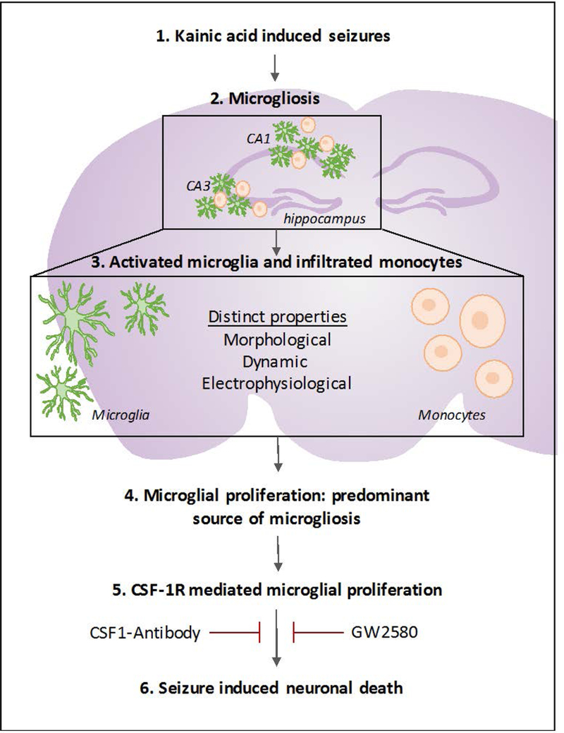
Figure 8. Seizure induced microgliosis in the hippocampus.

Schematic showing seizure induced microgliosis is comprised of morphologically and physiologically distinct activated resident microglia and infiltrated monocytes. Moreover, it illustrates that microglia, and not monocytes, proliferate in the hippocampus following seizures and that blocking microglial proliferation via antagonism of the colony stimulating factor 1 receptor may help reduce seizure-induced neuronal loss.
