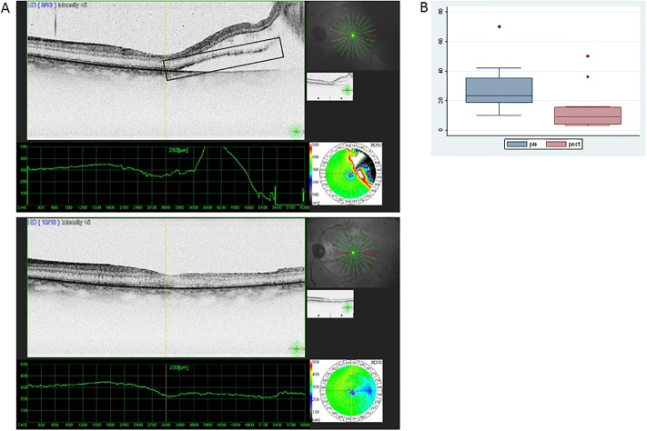
Fig 1. Presence of HR points in the retina before and after surgery for rRD.
(A) HR points are shown in the square box. OCT image from the retina when detached (image above) and after reattachment surgery (image below) showing absence of subretinal fluid and diminishing presence of HR points in the neuroretina. (B) Medians and Interquartile range of number of HR points counted on OCT images during the pre- (RD) and post- (reattachment) state (outliers being shown as separate dots).

