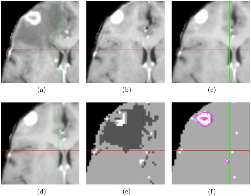
Fig. 1:
Example of registration and label map estimation results. Crosshairs mark the same position in the aligned space. (a) Source image. (b) Target image aligned using affine registration. (c) SNRR result. (d) Result using proposed method. Note the right ventricle is better aligned compared to the results in (b) and (c). (e) Label map showing increased (brighter) and decreased (darker) intensity in source compared to target. (f) Active tumor estimate for source image with hand-segmented tumors outlined in pink. The tumor estimate identifies the large tumor and the small metastases by the right ventricle (just left of vertical green line).

