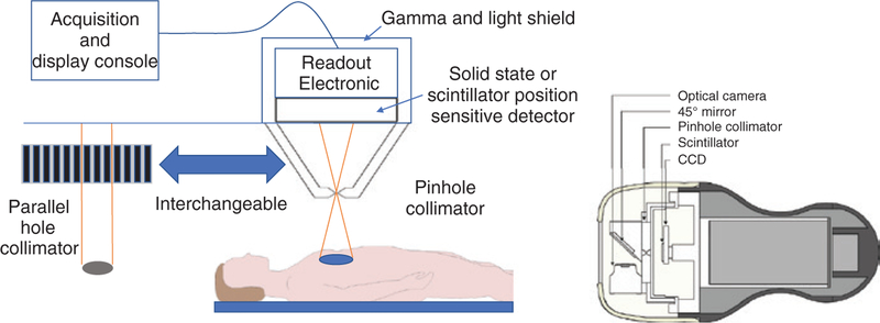
Figure 5. Intraoperative gamma imaging using a pinhole collimator.
A mirror at the entrance of pin-hole collimator is used to reflect optical photons to the CCD camera and produce coregistered images of optical and gamma photons. Reproduced with permission from [71].

