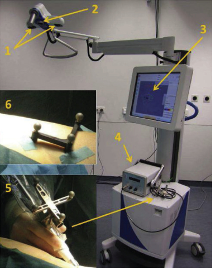
Figure 8. Surgical setup of a freehand SPECT system by Declipse (Surgieye, Munich, Germany).
The freehand 3D SPECT is composed of two calibrated infrared cameras (1) to determine accurate position of gamma probe in respect to the patient body using two tracking objects on patient (6) and gamma probe (5); and an optical camera (2) to record procedure flow and fuse the gamma distribution image to optical view of operation area in the display screen (3). A conventional gamma probe (4) generates gamma distribution data for 3D image creation before and after resection, as well as produces acoustic and visual feedback of gamma distribution during surgical procedures. Reprinted with permission from [119].

