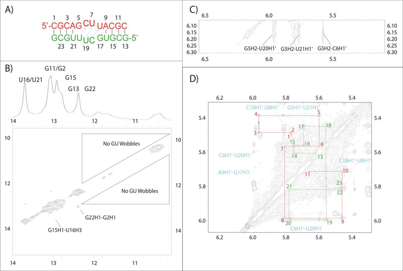
Figure 7.
(A) Predicted structure for GCUU. Arrows in cyan indicate residues that show cross-strand H1’-H1’ NOESY cross-peaks indicative of bifurcated GU pair formation. (B) 2D imino spectrum that reveals the structure outside of the internal loop formed as expected, despite the lack of GU wobble pairing in the loop region. (C) A NOE between G5’s amino and U20H1’ consistent with bifurcated pair formation. (D) H1’-H1’ walk region for both strands in red and green. The cross strand H1’-H1’ resonances that result from narrowing of the minor groove are shown in cyan.

