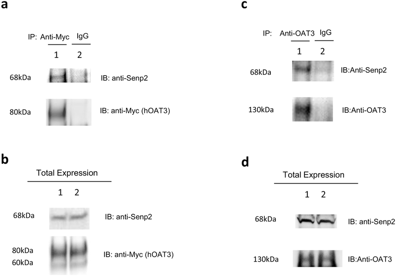
Fig. 9. Interaction between Senp2 and hOAT3.
(a) The Interaction of Senp2 with hOAT3 in COS-7 cells. Top panel: COS7 cells were co-transfected with hOAT3 and Senp2. Transfected cells were then lysed, and hOAT3 was immunoprecipitated with anti-Myc antibody or with normal mouse IgG (as negative control), followed by immunoblotting (IB) with anti-Senp2 antibody. Bottom panel: The same immunoblot from Fig. 9a, Top panel was reprobed by anti-Myc antibody. (b) Total expression of hOAT3 and Senp2. COS-7 cells were co-transfected with hOAT3 and Senp2 for 48h. Top panel: Cells were lysed, followed by immunoblotting (IB) with an anti-Senp2 antibody. Bottom panel: The same immunoblot from Fig. 9b, Top panel was reprobed by anti-Myc antibody. Line 1 and line 2 represent 2 samples. (c) The interaction of Senp2 with rOAT3 in rat kidney slice. Top panel: The kidney slices from rat were lysed, and rOAT3 was then immunoprecipitated with anti-OAT3 antibody or with normal mouse IgG (as negative control), followed by immunoblotting (IB) with anti-Senp2 antibody. Bottom panel: The same immunoblot as Fig. 9c, top panel was reprobed by anti-OAT3 antibody. (d) Total expression of rOAT3 and Senp2. Top panel: The kidney slices from rat were lysed, followed by immunoblotting (IB) with an anti-Senp2 antibody. Bottom panel: The same immunoblot from Fig. 9d, Top panel was reprobed by anti-OAT3 antibody. Line 1 and line 2 represent 2 samples.

