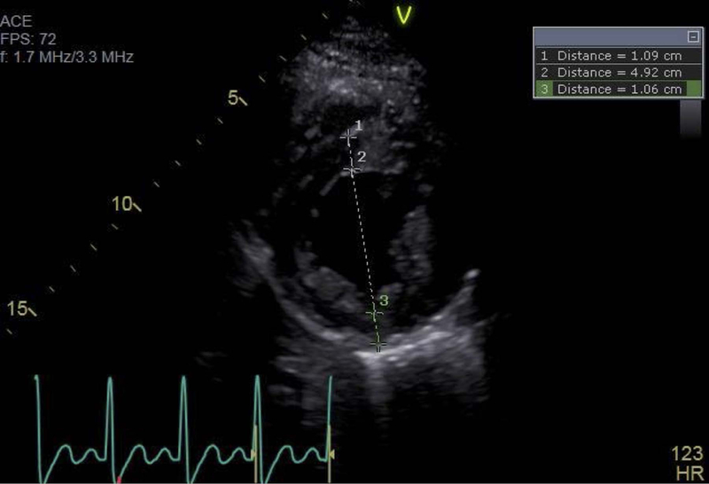
Figure 1:

Representative echocardiographic image, obtained at end diastole in the parasternal short axis view at the level of the mitral valve papillary muscles, demonstrating a typical relative wall thickness measurement. 1 = inter-ventricular septal thickness, 2 = left ventricular end diastolic internal diameter, 3 = left ventricular posterior wall thickness. In this example, RWT = (1.09 + 1.06)/4.92 = 0.44.
