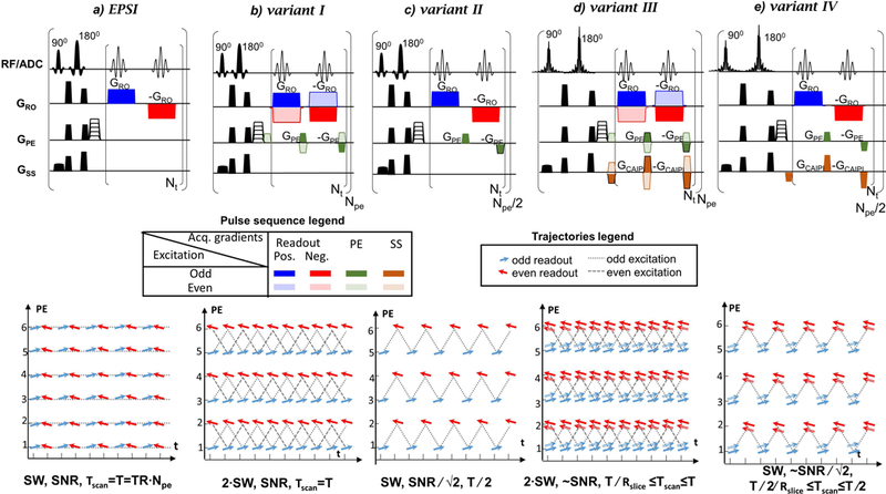
Figure 1:

Sequences (top) and PE/t trajectories (bottom) : a) EPSI, b)-e) consistent k-t space EPSI variants I-IV. The relative SW, SNR and total scan time (Tscan) for each sequence (a-e) are noted relative to E+O EPSI (a). The blue and red arrows correspond to positive and negative readout gradient lobes, respectively. The dual-band slice-selection has dual-arrows representing two slices. The odd/even excitations are marked differently as shown in the legends. Variant II and IV (d) and ( e) use only the odd excitations of the full schemes variants I and III (b) and (d), respectively. Rslice is the acceleration factor of the multi-band excitation (Rslice=2 for dual-band).
