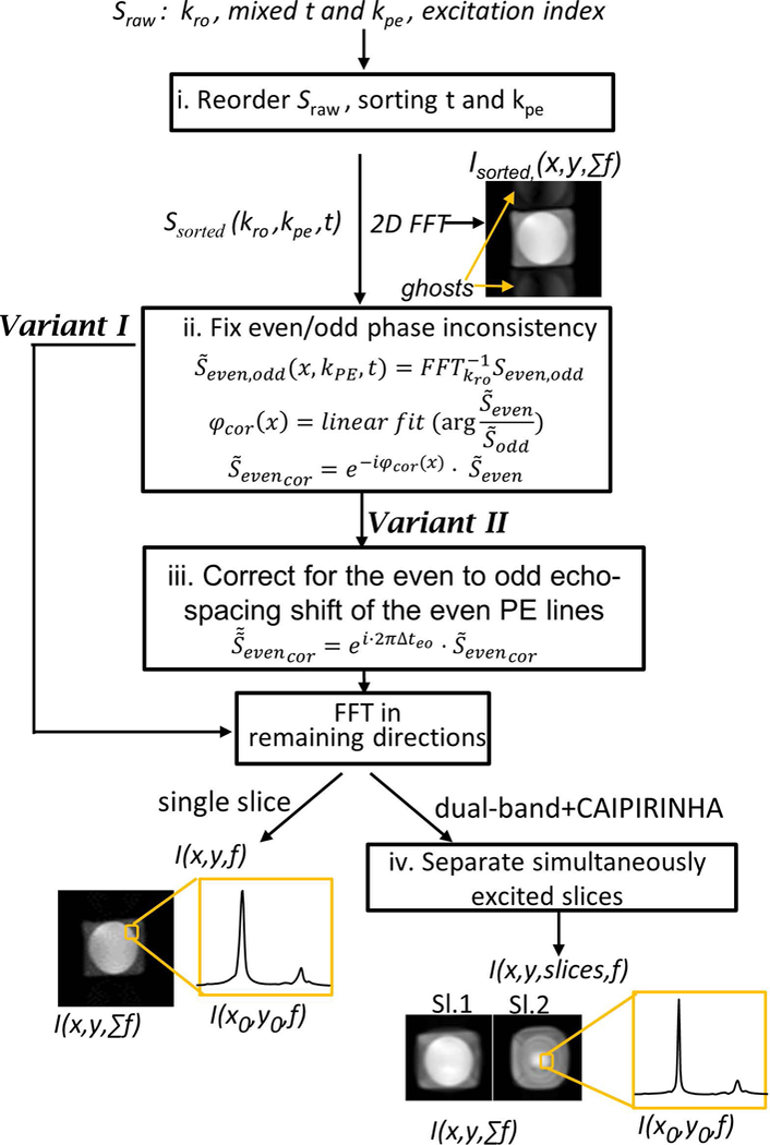
Figure 2:

Reconstruction steps employed for variants I and II and their dual-band (variants III, IV) extensions. Sraw is the input raw data signal and the I(x,y,f) for each slice is the spectral spatial final reconstructed output. The images after the first and last steps show representative results, demonstrating an image with ghosts that will appear after the first step and the final images that include spectroscopic and spatial information.
