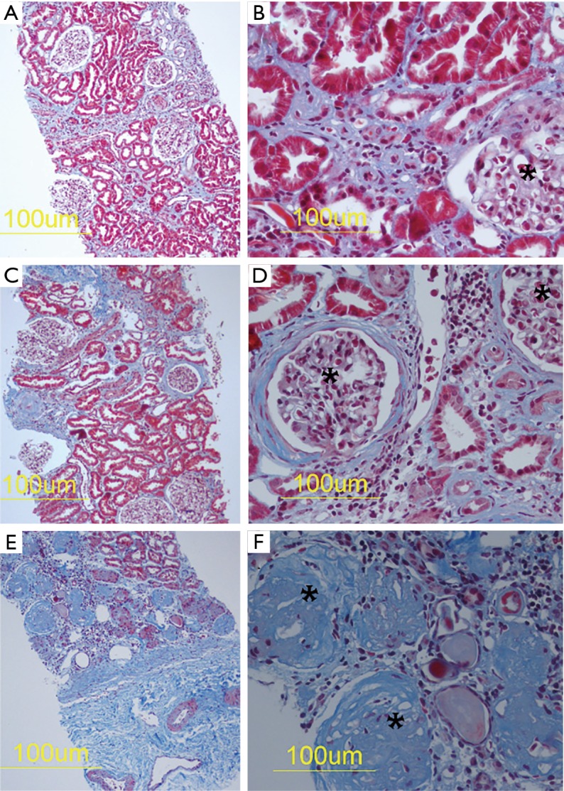
Figure 1.

Masson’s trichrome stain of renal biopsies. Masson’s trichrome histochemistry demonstrates mild (A,B), moderate (C,D) and extensive renal fibrosis (E,F) which is shown as blue in these photomicrographs. Glomeruli are marked with asterisks in (B,D,F). In (D), there is fibrotic thickening of Bowman’s capsule. In (F), there is marked glomerulosclerosis.
