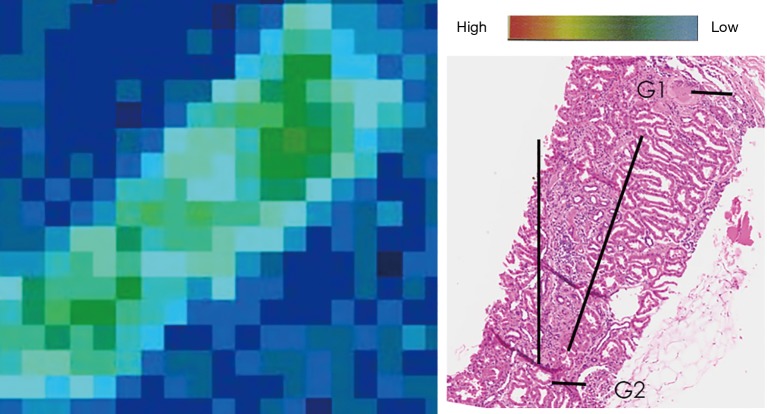
Figure 3.
SPring8 heatmap of cadmium and corresponding morphology of a Sri Lankan CKD sample. On the left is an example of SPring8 XRF heatmap imaging of cadmium in a kidney with moderate chronic kidney fibrosis. The hematoxylin and eosin histology staining on the right shows the morphology from the same biopsy area (×100). The color scale at the top right demonstrates XRF signal intensity for the heatmap of cadmium. There was no evidence of overlap of cadmium with fibrotic structures (G1 is a sclerotic glomerulus in comparison with G2 which is a healthy glomerulus; tubulointerstitial fibrosis is demarcated with the long straight black lines).

