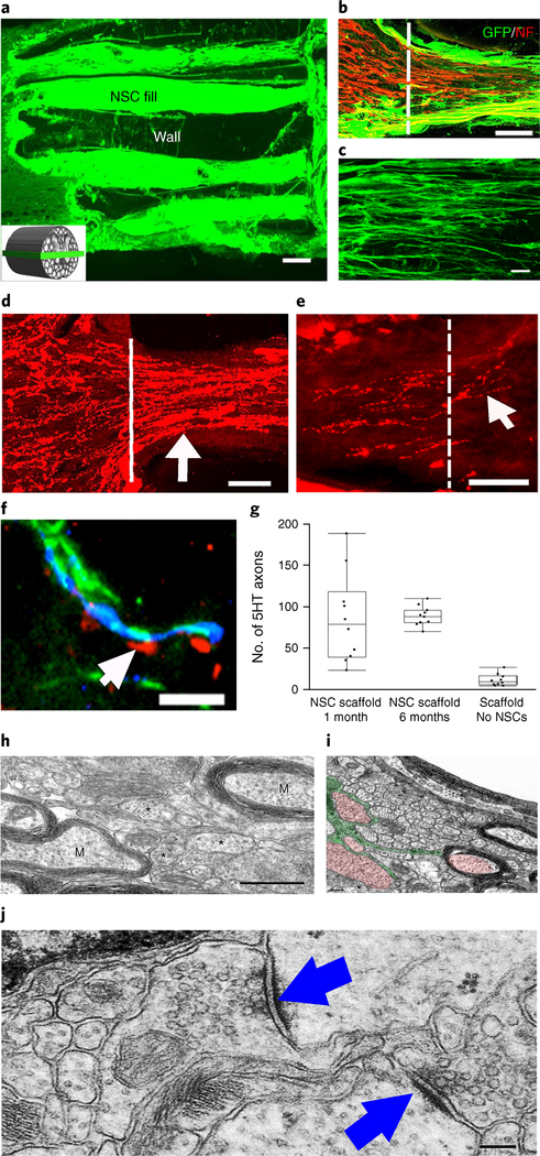
Fig. 3 |. Four weeks in vivo performance of NPC-loaded 3D-printed scaffold implants.
a, Channels are filled with GFP-expressIng NPCs. The inset schematic diagram indicates the orientation of the horizontal sections in all panels of this figure. b, The rostral entrance to the channel is penetrated by host axons (labeled for NF200 (NF)); the host axons are distinguished from graft-derived axons by the absence of GFP expression. c, Implanted GFP-expressing NSCs extend linear axons within the scaffold. Rostral is to the left and caudal is to the right. d, 5HT-labeled host serotonergic axons enter the NPC-filled channel from the rostral (left) aspect of the lesion and regenerate linearly in the channel (arrow). e, 5HT-labeled host axons exit the caudal aspect of the channel to regenerate into the host spinal cord distal to the lesion (arrow). The white line demarcates the exit from the caudal channel to the caudal spinal cord. f, 5HT host axons regenerating into scaffold channels form appositional contacts (arrows) with dendrites (MAP2) of implanted NPCs (labeled for GFP). g, Quantification of the mean number of 5HT axons reaching the caudal end of the scaffold (one-tail ANOVA P < 0.0322, post hoc Tukey’s), n = 10 animals. The boxes show the 25th-75th percentile range, and the center mark is the median. Whiskers show 1.5 times IQR from the 25th or 75th percentile values. h, At the ultrastructural level, axons of varying diameters (asterisks) are present within channels and many axons are myelinated (M). i, An ultrastructural image showing an oligodendrocyte (green) sending multiple processes to myelinate and ensheath axons (red). j, Synapses (arrows) form between axons within channels and the dendrites of implanted NPCs. The synapses are asymmetric with presynaptic boutons containing rounded vesicles. Scale bars, 200 μm (a), 50 μm (b), 10 μm (c,f), 100 μm (d,e), 500 nm (h), 0.2 μm (i), 200 nm (j).

