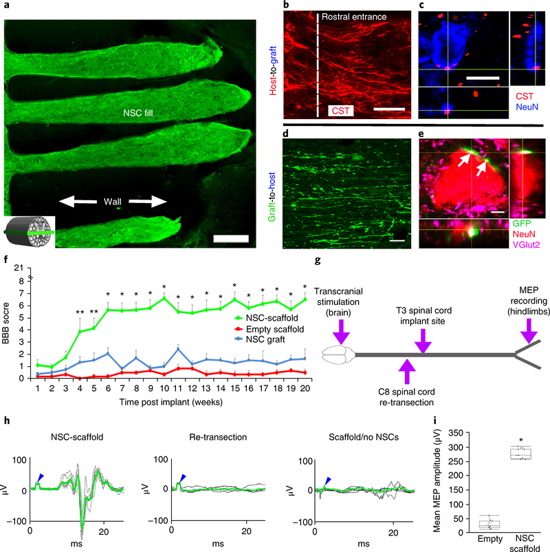
Fig. 4 |. Long-term in vivo studies of 3D-printed scaffolds loaded with NPCs.
a-e, Anatomy at 6 months post implant. a, The channels are structurally intact and filled with GFP-expressing NPCs. The inset schematic diagram indicates the orientation of the horizontal sections in all panels of this figure, with rostral to the left. b, Corticospinal axons enter the scaffold and extend linearly in a caudal direction. c, CST axons converge on a NeuN-labeled neuron inside the channel, forming bouton-like contacts with the soma. d, GFP axons extend out from the scaffold into the host white and gray matter caudal to the lesion. Ventrolateral white matter, 2 mm caudal to the lesion. e, NPC-derived GFP-labeled axons form excitatory contacts (VGlut2) on gray matter host neurons (labeled for NeuN) located 2 mm caudal to the lesion (white arrows). f-i, Behavioral studies. f, BBB motor scores after complete transection (repeated-measures ANOVA; **P < 0.0232, *P < 0.0008; mean±s.e.m, n = 10 animals). g, Schematic diagram of the electrophysiology study performed at 6 months post implant. Transcranial electrical stimulation is applied to the motor cortex in the brain and MEPs are recorded from the hindlimbs. h, Rats with 3D-printed, NPC-filled scaffolds exhibit MEP responses that are abolished by subsequent re-transection of the spinal cord immediately above the scaffold. Animals with empty scaffolds show no MEPs. The blue arrowheads mark stimulation artifacts. The green lines represent averages of several individual stimulations shown in black. i, The mean MEP amplitude is significantly greater in animals implanted with NPC-containing scaffolds. The boxes show the 25th-75th percentile range, and the center mark is the median. Whiskers show 1.5 times IQR from the 25th or 75th percentile values. (Student’s t-test; P < 0.0001, n = 10 animals). Scale bars, 250μm (a), 50μm (b), 10μm (c), 40μm (d), 5μm (e).

