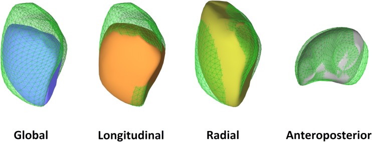
Fig. 3.
Representative case of a patient with pulmonary arterial hypertension (pulmonary vascular resistance 9.6 Wood units). The right ventricle is severely dilated (end-diastolic volume 234 mL) and global right ventricular function is decreased (ejection fraction 34%). By decomposing the motion of the right ventricle, a relatively preserved longitudinal function can be seen (longitudinal ejection fraction 17%), while radial (radial ejection fraction 8%) and anteroposterior shortening (anteroposterior ejection fraction 6%) is decreased. The basal predominance of right ventricular dysfunction is also notable

