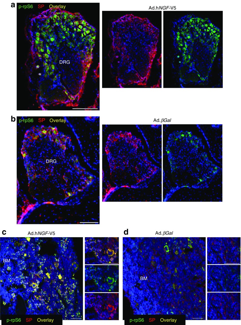
Fig. 2.
Gene therapy with NGF activates TrkA signalling in sensory neurons and bone marrow cells of diabetic mice. (a, b) Representative immunofluorescence images showing that, 16 weeks post gene transfer, a higher number of substance P-positive sensory neuron bodies display phosphorylation of rpS6 in the DRG of diabetic mice receiving V5-tagged Ad.hNGF (a) compared with diabetic mice receiving Ad.βGal (b). Scale bar, 100 μm. (c, d) Representative immunofluorescence images showing that, in bone marrow of hNGF-treated mice, a higher number of cells are characterised by phosphorylation of rpS6 (c) compared with control mice receiving βGal (d). Scale bar, 50 μm. In all images, phospho-rpS6 is shown in green, substance P in red. DAPI, in blue, identifies nuclei. n = 3 mice, Ad.hNGF and n = 3 mice, Ad.βGal. BM, bone marrow; SP, substance P

