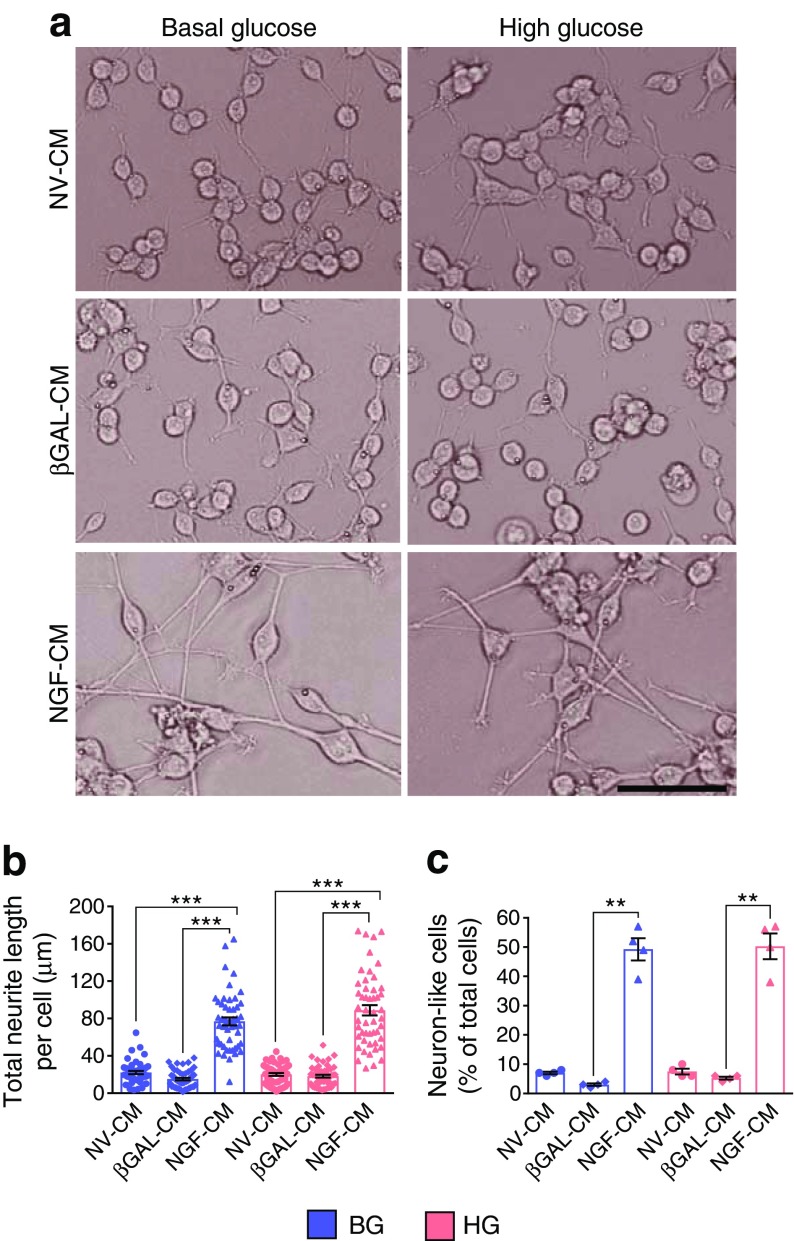
Fig. 5.
hNGF induces neuronal differentiation of PC12 cells in vitro. (a) Bright-field images showing that PC12 cells differentiate into neuron-like cells in the presence of hNGF for 3 days, in either basal- or high-glucose environments (for the schematic of the experiment, please see Fig. 4b). Scale bar, 50 μm. (b) Graph showing the total neurite length per cell (n = 50 cells per group assessed in four different imaging fields). (c) Graph showing the percentage of neuron-like cells, defined as cells presenting at least one axon longer than the cell body (n = 4 different imaging fields per group, for a total of n = 250–300 cells assessed per group). Data are expressed as means ± SEM. **p < 0.01 and ***p < 0.001 between groups, as indicated. Blue bars/symbols, basal glucose; pink bars/symbols, high glucose. BG, basal glucose; CM, conditioned medium; HG, high glucose; NV, non-virus

