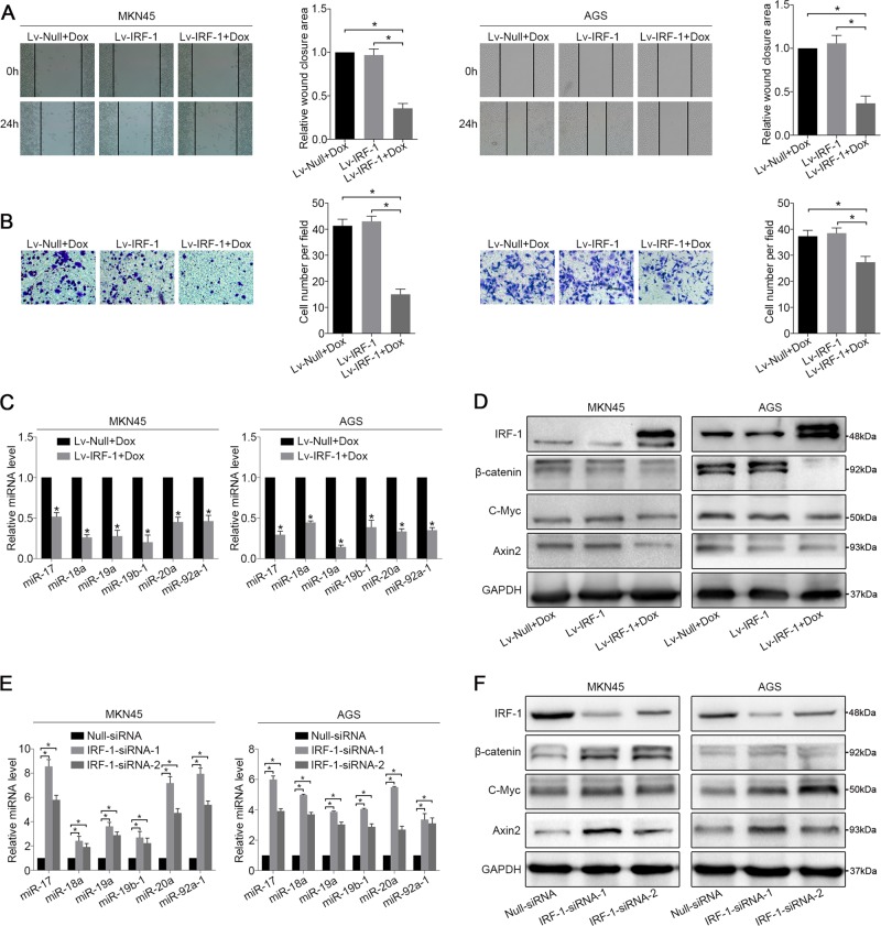
Fig. 3. IRF-1 regulates MIR17HG expression.
a Wound-healing and b migration assays of Lv-IRF-1 Dox-treated, Lv-IRF-1 Dox-untreated, and Lv-Null Dox-treated MKN45 and AGS cells were performed. c After 48 hours of Dox induction, the expression of six polycistronic miRNAs derived from MIR17HG in the MKN45 and AGS cell lines transfected with Lv-IRF-1 and Lv-Null was analysed by qRT-PCR. d After 48 hours of Dox induction, IRF-1, β-catenin, C-Myc and Axin2 expression in the MKN45 and AGS cell lines transfected with Lv-IRF-1 and Lv-Null was analysed by western blot analysis. e qRT-PCR analysis of the expression of six polycistronic miRNAs derived from MIR17HG in the MKN45 and AGS cell lines after IRF-1 knockdown. f Western blot analysis of IRF-1, β-catenin, C-Myc and Axin2 expression in MKN45 and AGS cell lines after IRF-1 knockdown. All the above experiments were independently performed in triplicate (N = 3). *P < 0.05, as determined by paired Student’s t test. The data a, b, c and e are presented as the means ± SDs

