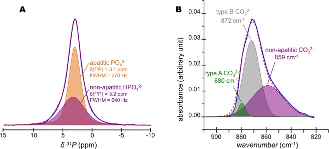
Figure 5.
Quantification of HPO42− and CO32− ions present in bone mineral. (A) Quantitative 31P single pulse (SP) magic angle spinning (MAS) solid-state Nuclear Magnetic Resonance (ssNMR) spectrum of a fresh 2-year-old sheep bone tissue sample (blue line) and its corresponding fitting (red dashed line) with two peaks. Those two peaks, whose lineshape and linewidth were revealed in Fig. 4B, correspond to the PO43−-containing internal crystalline core in the form of hydroxyapatite (orange peak) and the HPO42−-containing non-apatitic environments in the form of an amorphous surface layer (purple peak). (B) Fourier Transform-Infrared (FT-IR) spectrum of the ν2(CO3) vibration mode for a 2-year-old sheep bone tissue sample (blue line) and its corresponding fitting (red dashed line). Type B CO32− ions occupy the PO43− sites within the hydroxyapatite’s crystal lattice; type A CO32− ions occupy the OH− sites within the hydroxyapatite’s crystal lattice; whereas non-apatitic CO32− are present within the amorphous surface layer that coats bone mineral particles.

