Figure 2.
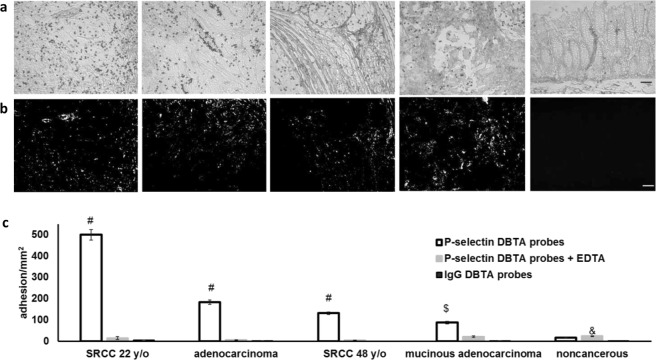
DBTA signal is specific, quantifiable, and discernible from SBTA. (a) P-selectin DBTA probes adhesion at 0.50 dyne/cm2 to colon tissue (from left to right: SRCC T4N1M0 22 y/o, adenocarcinoma T4N0M0, SRCC T4N1M0 48 y/o, mucinous adenocarcinoma T4N2M0, noncancerous colon tissue). DBTA probes appear as black circles. (b) P-selectin SBTA conducted on serial sections of the tissues examined in (a) with DBTA revealed detection of ligands that are not in complete agreement with the purported functional ligands detected via DBTA (a). Order of tissues is the same as (a). (c) P-selectin DBTA probe adhesion to colon tissue was specific and significantly greater than control probes. DBTA probes were perfused at 250,000 probes/mL and 0.50 dyne/cm2. Specificity of interaction was confirmed using 10 mM EDTA (divalent cation chelator, Ca2+ is required for selectin/selectin-ligand binding) and hIgG DBTA probes as negative controls. Data shown are mean adhesion ± SD of three technical replicates and are representative of independent experiments conducted on tissue sections from >10 independent cases of colon cancer and >3 independent noncancerous cases. #P < 0.001, $P < 0.01, and &P < 0.05, compared to all others intragroup. Scale bar = 100 µm.

