Figure 4.
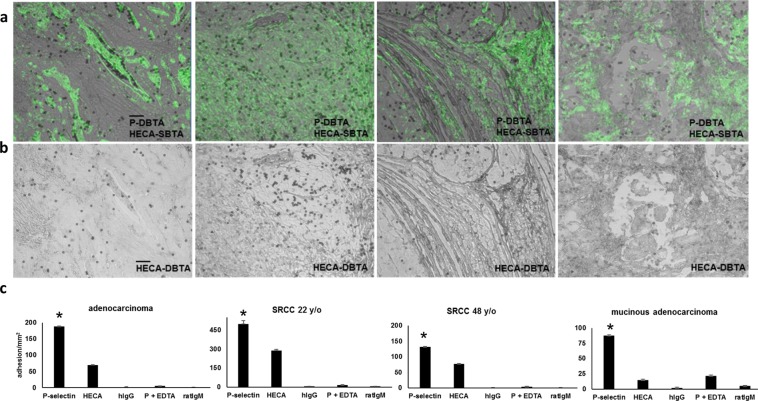
The presence of sLeX/A does not dictate a functional P-selectin ligand. (a) SBTA with the HECA-452 monoclonal antibody (green pseudocolor), which detects both sLeX and sLeA, has been combined with an image showing the adhesion patterns of P-selectin DBTA probes (Fig. 2). HECA-452 SBTA was conducted on serial sections. For the adenocarcinoma and SRCC 22 y/o tissues, but not the SRCC 48 y/o and mucinous adenocarcinoma tissues, there was an agreement between P-selectin DBTA and SBTA with HECA-452 in which the regions of tissue that express sLeX or sLeA mediate adhesion to P-selectin DBTA probes. However, with regard to the SRCC 48 y/o and mucinous adenocarcinoma tissues, not all regions that express sLeX or sLeA, as detected by HECA-452 SBTA, appeared to mediate binding to P-selectin DBTA probes (Supplementary Videos S1, S4, and S7). (b) HECA-452 DBTA probes (microspheres coated with HECA-452) adhered to tissue at 0.50 dyne/cm2 (Supplementary Videos S9 and S10). (c) The greatest amount of adhesion to the tissue occurred with the P-selectin DBTA probes. Probes were perfused at 250,000 probes/mL and 0.50 dyne/cm2. Specificity of interaction was validated using hIgG DBTA probes, 10 mM EDTA, and rIgM DBTA probes as controls. Data shown are mean adhesion ± SD of three technical replicates and are representative of independent experiments conducted on tissue sections from >3 independent cases of colon cancer. *P < 0.0025, compared to all others intragroup. Scale bar = 100 µm.

