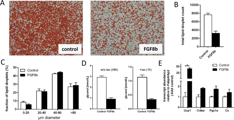
Figure 4.
Fgf8b interferes in adipocyte differentiation. (A) The number of mature, lipid-laden adipocytes is reduced at the end of differentiation in a medium containing Fgf8b (125 ng/ml) versus control treated cells (con). Orange colour indicates automatic detection of a lipid droplet by an image analysis algorithm. (B,C) Final lipid droplet number is decreased (B), while size distribution is similar (C) after Fgf8b treatment. (D) Unstimulated (left panel) and isoproterenol-stimulated (iso, right panel) lipolytic rate determined by glycerol release are both decrease after differentiation in the presence of Fgf8b. Bars depict mean values ± SD, n = 3–4. (E) Transcript abundance of brown/brite adipocyte markers UCP1, Cidea and Pgc1a was increased by Fgf8b when normalized to the marker of terminal adipocytes differentiation AP2/Fabp4. Ucp1, uncoupling protein 1; Cidea, cell death-inducing DNA fragmentation factor alpha-like effector A; Pgc1a, peroxisome proliferator-activated receptor gamma, coactivator 1 alpha; Cs, citrate synthase; bars are mean values ± SD, n = 3. Stars indicate a significant difference (p < 0.05, Sidak’s multiple comparisons test).

