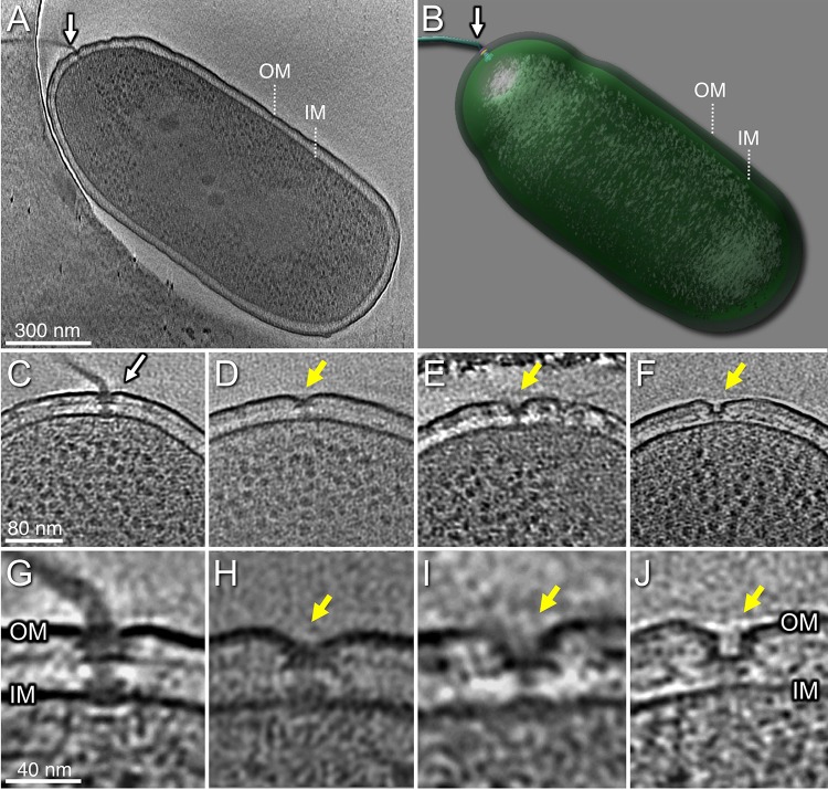
FIG 1.
Tomograms of P. aeruginosa PAK cells show the intact polar flagellum and subcomplexes. (A) One slice from a whole-cell tomogram shows a single polar flagellum. (B) A surface rendering of the tomogram shown in panel A. (C) A slice from one tomogram shows the intact flagellar motor. (D) A slice from one tomogram shows a flagellum lacking the hook and filament. (E) A tomogram slice shows a flagellum without the rod, hook, and filament. (F) A tomogram slice shows a novel structure with a large pore in outer membrane (OM). (G) A zoomed-in view of the flagellar motor shows major components embedded in OM and inner membrane (IM). (H) A zoomed-in view of the motor in panel D. (I) A zoomed-in view of the motor in panel E. (J) A zoomed-in view of the outer membrane pore in panel F.

