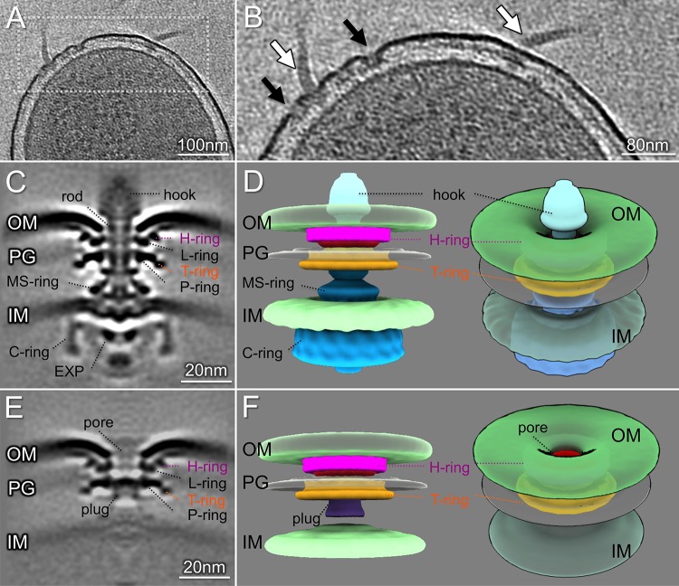
FIG 2.
Structures of the P. aeruginosa flagellar motor and subassembly. (A) A representative slice of a cell pole reconstruction from P. aeruginosa ΔfleN showing multiple flagella. (B) A zoomed-in view of the slice shown in panel A. Flagellar motors and hooks are indicated by white arrows. The flagellar outer membrane complexes (FOMCs) are shown by black arrows. (C) A slice of an averaged structure of the intact flagellar motor. Note the striking curvature of the outer membrane. (D) Side and tilted views of the three-dimensional (3D) surface rendering of the motor show the curvature of the outer membrane and the FOMC. (E) A slice of an averaged structure of the FOMC. (F) A 3D surface rendering of the FOMC. The novel “plug” is colored purple. A tilted view of the 3D surface rendering shows the membrane pore in the FOMC. EXP, export apparatus; OM, outer membrane; PG, peptidoglycan layer; IM, inner membrane.

