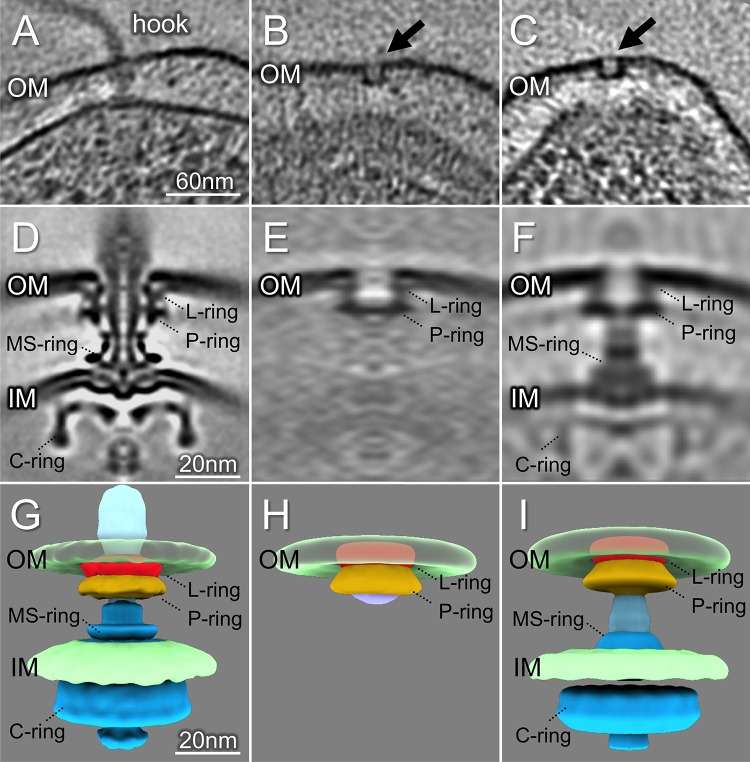
FIG 3.
Intact flagellar motor, FOMC, and a subcomplex revealed in S. Typhimurium minicells. (A) A representative tomogram slice of an S. Typhimurium minicell shows a complete flagellar motor. (B, C) Two representative slices show FOMCs in S. Typhimurium. (D) A slice of a subtomogram average of the complete S. Typhimurium flagellar motor. (E) A slice of the averaged structure of the FOMC. (F) One class average shows the FOMC assembly aligned with the MS-ring in the absence of a distal rod. (G) Surface view of the intact flagellar motor. (H) A 3D surface rendering of the FOMC structure. (I) Surface rendering of a subcomplex in which the MS-ring is associated with the FOMC.

