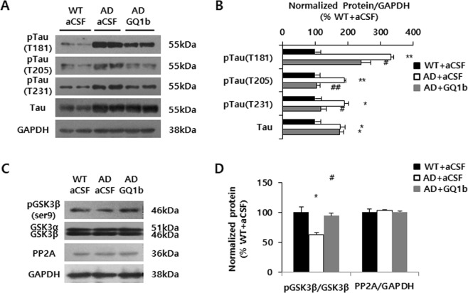
Figure 5.
GQ1b decreases tau phosphorylation accompanied by increased levels of phospho-GSK3β in 3xTg-AD mice. (A) Western blotting and (B) its quantification revealed that GQ1b administration into the hippocampus of 3xTg-AD mice significantly reduced phospho-tau (T181, T205, T231) levels compared to aCSF-infused 3xTg-AD mice. (C) Western blotting and (D) its quantification showed that GQ1b infusion rescued the decrease in phospho-GSK3β levels without affecting expression of a representative tau phosphatase, PP2A. Western blotting band intensity was quantified by densitometry analysis on pTau, Tau, pGSK3β, GSK3β, PP2A, and GAPDH bands. Western blots shown represent typical results from three independent experiments, and the graphs show data from three independent experiments and are expressed as mean values ± SD. *p < 0.05, **p < 0.01 vs. WT + aCSF group, #p < 0.05, ##p < 0.01 vs. AD + aCSF group.

