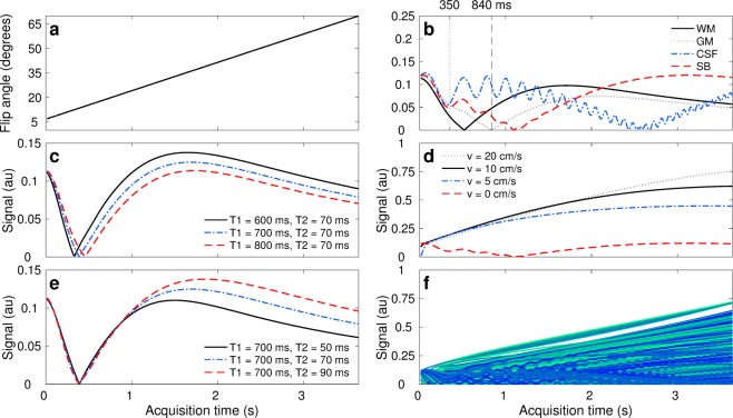
Figure 2.
Parameter encoding scheme and signal dynamics in the presence of flow. (a) Encoding strategy given by linear flip angles variations. (b) Signal evolution for each tissue class: grey matter (GM), white matter (WM), cerebrospinal fluid (CSF), and stationary blood (SB). The sequence has been designed to have distinct signal evolutions for each class; where SB contrast discriminates well from the others towards the end of the sequence. Our proposed reconstruction framework also enables the visualisation of tissue dynamics through time, where e.g. WM/GM tissue contrasts at 350 and 840 ms are inverted as they recover from the inversion pulse with different T1 relaxation times. (c,e) Representation of parameter encoding with transient-state signal evolutions. Tissues with shorter T1 times experience their inversion first in time (solid black line in c) whereas tissues with longer T2 times produce larger stimulated echoes and thus less signal decay (dashed red line in e). (d) Hyperintensities due to fast flowing spins with 2 mm slice thickness. The thinner the imaging slice or the faster the flow, the more pronounced flow effects become. (f) Cohort of signals obtained by sampling the prior probability distribution of stationary and non-stationary tissues. As signals exhibit correlation in space and time, lower dimensional constraints can be imposed for image reconstruction. The proposed design scheme satisfies maximal contrast conditions and optimal parameter encoding in 3.66 seconds per imaging slice.

