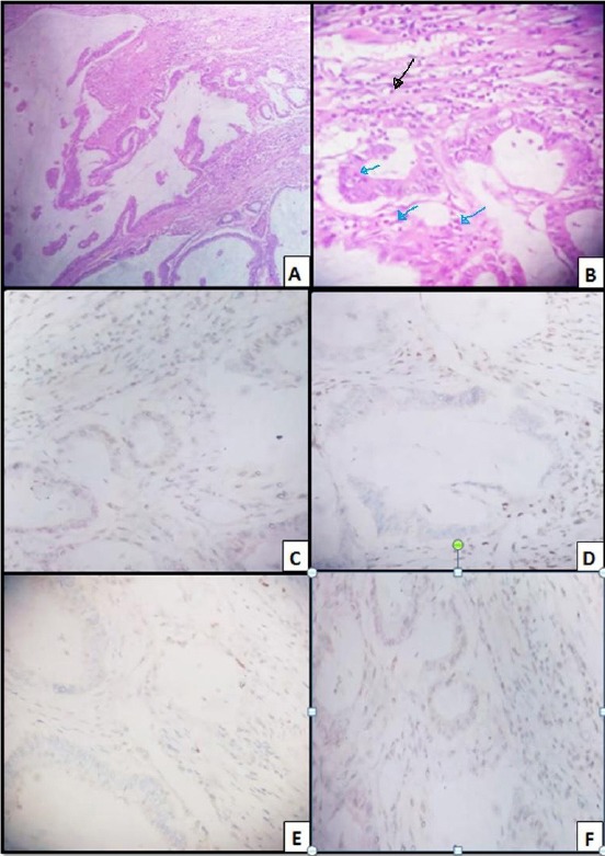
Figure 2.

Colonic mucinous carcinoma; A) H&E (x 100), Pools of mucin entangling malignant glands; B) H&E (x 400), TIL (blue arrows); PTL (black arrow); C) MLH 1; Weakly positive nuclear staining of tumor cells (x 400), with positive lymphocytes, internal control; D) MSH2, Negative nuclear staining of tumor cells (x 400). with positive lymphocytes, internal control; E) MSH6, Negative nuclear staining of tumour cells (x 40); F) PMS 2, Weakly positive staining of tumour cells (x 400)
