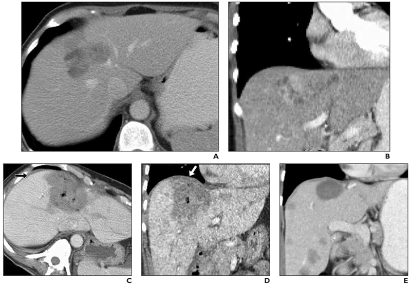
Fig. 3—

59-year-old woman with 6-cm metastatic uterine sarcoma in segment 8 of liver.
A, Preprocedure IV contrast-enhanced CT scan.
B, Preprocedure coronal IV contrast-enhanced CT scan.
C, Immediate postprocedure IV contrast-enhanced CT. Large ablation zone contacts diaphragm anteriorly. Note thin layer of fluid (arrow) as result of hydrodissection (450 mL of 5% dextrose in water) that separates liver capsule from diaphragm.
D, Immediate postprocedure coronal IV contrast-enhanced CT. Large ablation zone extends to hepatic dome. Note minimally increased thickness of diaphragm adjacent to ablation zone (arrow). Patient experienced mild shoulder pain (3/10 maximum) after procedure.
E, Follow-up CT 5 months later shows marked decrease in size of ablation zone, no evidence of diaphragm hernia or residual thickening, no local tumor progression, but new hepatic metastases.
