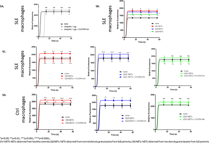
Figure 5. Extracellular ubiquitin and NETs increase calcium flux in macrophages.
Monocyte-derived macrophages from SLE subjects and healthy controls were obtained. They were stimulated with recombinant ubiquitin (1 μg) (A) or with 50 μg of NETs from healthy controls, lupus normal density granulocytes and lupus LDGs (B-D). In addition, a CXCR4 (extracellular ubiquitin receptor) inhibitor was used (A,C,D). Calcium flux was measured by Fluo-4 NW Calcium Assay Kit in all cases (5 independent experiments). Calcium flux was enhanced by recombinant ubiquitin (A) and by NETs (B-D), and significantly decreased when a CXCR4 inhibitor was added. There was also a differential calcium influx in response to NETs from healthy controls, SLE patients and LDGs. *p<0.05; ** p<0.01; ***p<0.001; ****p<0.0001; n.s.=non significant.

