
Figure 6. SLE macrophages internalize NETs and synthesize cytokines in response to NET internalization.
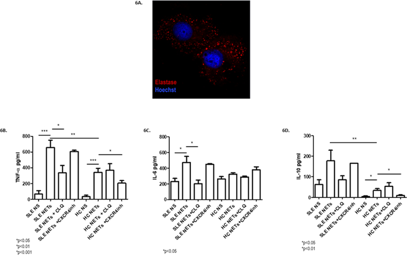
Monocyte-derived macrophages from SLE and healthy subjects were incubated with 50 μg of NDG NETs from SLE subjects for 6 hours (N=18). A) Immunofluorescence was performed and analyzed by confocal microscopy. Red represents neutrophil elastase and blue represents DNA. A representative image is shown, demonstrating internalization of neutrophil elastase from NETs by macrophages. B-D) Supernatants were obtained, and cytokine levels (TNF-α, IL-6 and IL-10) were measured by ELISA. Boxes show the pooled data (mean and SEM) of each cytokine assessed in non-stimulated (NS) SLE and control macrophages, macrophages stimulated with NETs, macrophages stimulated with NETs after chloroquine pretreatment, and macrophages stimulated with NETs after treatment with a CXCR4 inhibitor (AMD3100). There was a higher TNF-α and IL-10 synthesis in SLE macrophages compared with controls when they were stimulated with NETs. When SLE macrophages were pretreated with chloroquine, IL-6 and TNF-α release significantly decreased. After CXCR4 inhibition, there was a decrease in TNF-α and IL-10 synthesis in control macrophages; *p<0.05; ** p<0.01; ***p<0.001; n.s.=non significant.