
Fig. 1.
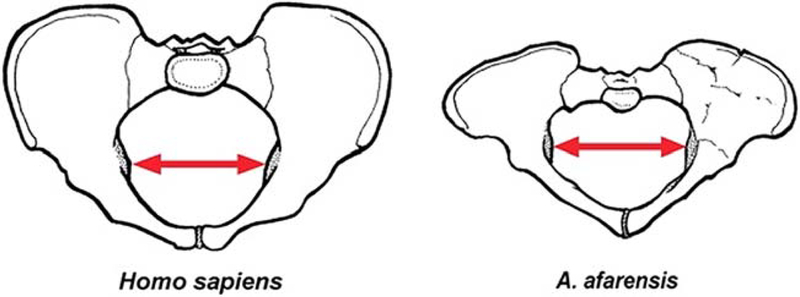
Pelvic dimensions in female modern Homo sapiens (left) and female Au. afarensis specimen A.L. 288–1 (“Lucy,” right). The two pelves are shown in in superior/anterior view and drawn to the same scale. The red arrows indicate bi-acetabular breadth, which is very similar in the two species despite the great difference in overall body size. Modified from Rosenberg and Trevathan (2002).