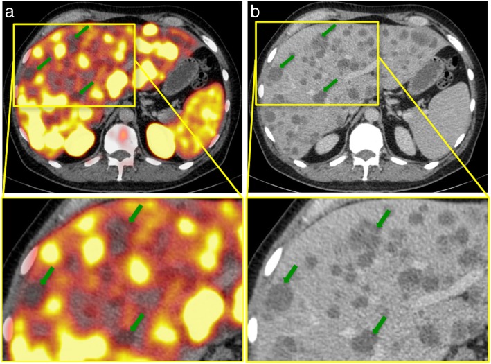
Fig. 2.
Example of 68Ga-PSMA-negative liver metastases in a PC patient with a recurrent adenocarcinoma. a, b: 68Ga-PSMA-PET/CT of a 54-year-old patient with a recurrent adenocarcinoma of the prostate and disseminated lymph node, bone, and hepatic metastases. After the initial diagnosis in 2011, the patient had received a radical prostatectomy and undergone chemotherapy as well as androgen-deprivation therapy. The serum PSA was 4962.0 ng/ml at the time of examination; the initial GS was 4 + 5. The PET/CT (a) illustrates disseminated, PSMA-negative liver metastases, with SUVmax-values up to 4.2 (liver background 9.5). Green arrows point to examples of liver metastases in segments IVa and V. In CE-CT (b), liver metastases appear hypodense compared to the liver. GS Gleason score, PSA prostate-specific antigen, SUVmax Maximum standardized uptake value

