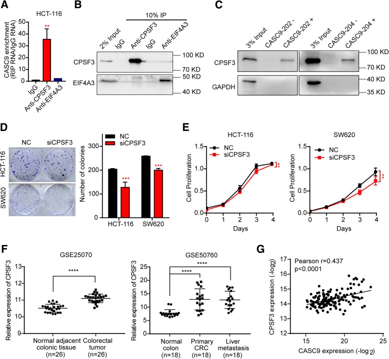
Fig. 6.
CPSF3 interacts with CASC9 and regulates CRC cell growth. a CASC9 RNA was measured by RT-qPCR in IgG control-, CPSF3-, and EIF4A3-RIP samples. b Validation of CPSF3- and EIF4A3-immunoprecipitation efficiency by western blotting. c Interaction between CASC9–202 or CASC9–204 and CPSF3 was validated by RNA-protein pull-down and western blotting. GAPDH was used as a negative control. d, e HCT-116 and SW620 cells were transfected with siRNAs targeting CPSF3 (siCPSF3) or negative control (NC). Cell proliferation was determined by colony-formation assay (d) and, at the indicated time points, by MTS assay (e). f Scatter plot showing that the expression of CPSF3 was significantly upregulated in CRC tissues as compared to adjacent normal colon tissues in the GSE25070 (n = 26, left) and GSE50760 (n = 18, right) datasets. g Correlation between CASC9 and CPSF3 expression in 155 human CRC tissues. Expression data for CASC9 and CPSF3 were downloaded from the lncRNAtor database. The data are presented as the mean ± s.d. **P < 0.01, ***P < 0.001, ****P < 0.0001 by Student’s t-test (a, d), two-way ANOVA (e), or Wilcoxon signed-rank test (f)

