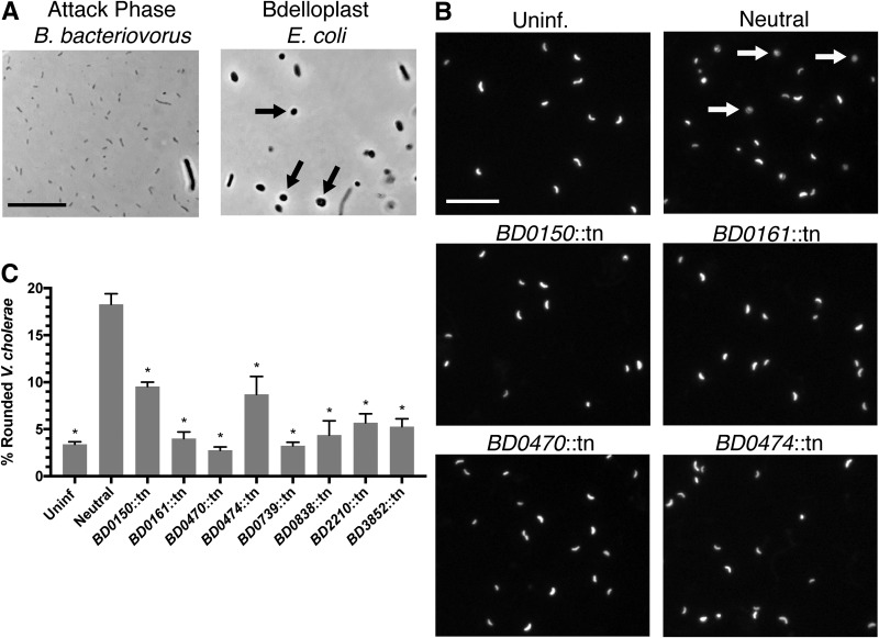
FIG 4.
Identification of B. bacteriovorus mutants with defects in prey rounding by Tn-SphereSeq. (A) Microscopy images of attack-phase B. bacteriovorus and E. coli bdelloplasts isolated by differential centrifugation for Tn-SphereSeq. Arrows indicate bdelloplasts. (B) Fluorescence images of GFP-expressing V. cholerae 3 h following infection with B. bacteriovorus at an MOI of 1. Arrows indicate bdelloplasts. Scale bar = 10 μm. (C) The percentage of rounded V. cholerae cells was calculated by analyzing images by Matlab for roundness (eccentricity) of three biological replicates. Significance was determined by comparing each strain’s rounding percentage to that of the neutral control. *, P < 0.0001 (ANOVA with Dunnett’s multiple-comparison test).

